

Viewing 3D Structures
ProtSpace integrates with AlphaFold to display 3D protein structures alongside your embedding visualization.
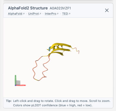
How It Works
When you select a protein with a UniProt accession:
- The structure viewer appears in the sidebar below the legend
- A row of links to AlphaFold Database, UniProt, InterPro, and TED appears under the title - click them anytime, each opens in a new tab
- The AlphaFold structure file is fetched directly from the AlphaFold Database API; the 3D-Beacons API is used only to look up the model page link
- Optional TED domain annotations are requested from
https://alphafold.ebi.ac.uk/api/domains/{accession}; the structure still loads if annotations are unavailable
Supported Structures
Currently, ProtSpace supports AlphaFold structures only. PDB experimental structures are not yet integrated.
Confidence and Domain Coloring
Structures initially use predicted Local Distance Difference Test (pLDDT) confidence scores, the same scheme used on the AlphaFold Database. Regions in blue are high-confidence, yellow moderate, and red low-confidence. This helps you quickly spot which parts of the model are more reliable.
Use the Color by control to switch between pLDDT and TED domains without reloading the structure. TED mode assigns a consistent categorical color to every segment of a domain and shows residues without a TED assignment in gray. The TED domains option is disabled when valid annotations are unavailable for the selected protein.
Viewer Controls
| Action | Effect |
|---|---|
| Color by pLDDT/TED domains | Switch the loaded structure's color theme |
| Left drag | Rotate the structure |
| Right drag | Pan the view |
| Scroll | Zoom in/out |
| Double-click | Reset the view |
When Structures Aren't Available
Not all proteins have AlphaFold structures. When no structure is found, the viewer displays:
"No 3D structure was found for <Protein ID>"
Next Steps
- Exporting Results - Save your findings
- FAQ - Common questions